Bacterial biofilms in medicine
Project 1: Superbugs in hospital environments
Understanding and eradicating an important source of nosocomial infection
Introduction
Between 6-10% of all hospitalised patients develop nosocomial infections, which cause increased morbidity and mortality and result in increased healthcare costs. The environment is one important source of cross-infection and we have recently shown that most surfaces in hospitals are contaminated with biofilms which can contain multi-antibiotic resistant organisms (MRO). The risk of obtaining a nosocomial infection is increased by an average of 73% if the previous patient occupying that room had a MRO. The role of biofilms in maintaining MRO viability and transmission of infection is unknown and will be explored in this study.
Hypothesis:
- Persistent exposure to antibiotics and disinfectants in the ICU induces biofilm growth, which subsequently promotes maintenance of MROs;
- Infection of multiple patients, over time, with the same strain of MRO is due to the periodic release of free swimming bacteria from these biofilms;
- Current cleaning methods and auditing do not deal with dry surface biofilms
Aims:
- Determine if MRO persist over time as biofilms and contribute to nosocomial infection.
- Compare environmental biofilm formation in the face of detergent cleaning compared to detergent cleaning plus chlorine disinfection with respect to MRO persistence.
- Show that quadruple monitoring of environmental surfaces with real-time feedback to cleaners, combined with new generation cleaning, will decrease environmental contamination, remove demonstrable MROs and biofilms from the hospital environment and decrease patient acquisition of MROs.
Research plan:
Aims 1 and 2. Patients admitted to intensive care units in Australia and Scotland will be screened on admission, weekly and on ICU discharge for MRSA, vancomycin resistant enterococci (VRE) and multidrug resistant gram-negative bacilli (MDRG). Line-associated bacteraemia, S. aureus bacteraemia and MRO data will be collected. All MRO patient strains will be collected and stored at -80oC. Patient demographics, date and bed number(s) will be entered into a database. Those patients with no prior history of MRSA, who had been on the ward for >48 hours and have laboratory isolated MRSA from their clinical samples will be regarded as a new MRO acquisition. Collection of environmental samples will be conducted after routine cleaning and disinfection. Ten high "hand-touch" sites will be screened weekly using chromogenic dip slides. 5 environmental samples for biofilm analysis will be collected monthly for 6 mths from 10 patient bed areas and the nurses' station before being divided into sections and processed by culture, quantitative real-time PCR, next generation sequencing, confocal laser scanning microscopy and scanning electron microscopy. Patient, environmental planktonic and biofilm bacterial strains will be subjected to molecular characterisation by whole genome sequencing. We will correlate geographic and time relationships between environmental, biofilm and patient strains to determine the contribution of the environment, in particular biofilms, strains to nosocomial infection.
Aim 3. Validation that cleaning has removed pathogenic organisms from the environment is important for infection control. Cleaning validation tools include visual inspection, fluorescence marker testing, ATP sampling, and microbial sampling for culture and biofilm demonstration. We will use failure mode and effects analysis to identify and grade each failure point in cleaning and the audit process, the frequency of this failure occurring, the effect of this failure, and whether this failure can be detected during normal protocols.
Enquires:
Associate Professor Karen Vickery
Email: karen.vickery@mq.edu.au
Project 2: Bacterial biofilm and breast implant associated anaplastic large cell lymphoma (ALCL)
Introduction
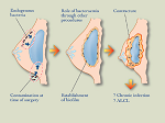
A recent association between breast implants and the development of ALCL has been observed. Both animal and human studies have shown bacterial biofilm infection causes capsular contracture and that a higher bacterial load produces chronic immune activation around breast implants. A number of reported observations about ALCL point to underlying biofilm infection as a potential cause.
 infection, inflammation and subsequent lymphomagenesis.
- The higher association of textured implants with ALCL which is consistent with our finding that textured implants support a higher bacterial load
- The late onset of ALCL following insertion of breast implants allowing underlying inflammation from bacteria to stimulate lymphoproliferation
- Late seroma, the commonest clinical presentation of ALCL is associated with both textured implants and biofilm
- Preliminary data show a significant association between T cell hyperplasia and T cell activation related to bacterial infection and texturisation of breast implants in both pigs and humans.
- The known association between bacterial infection and lymphoma in both humans (H Pylori and MALT gastric lymphoma) establishes a pathway from bacterial
Hypothesis:
- The trigger for both reactive lymphoproliferation and potential malignant transformation may be underlying chronic bacterial biofilm infection of breast implants.
- The microbiome associated with ALCL is significantly different from the microbiome associated with capsular contracture.
Aims:
- Assess the human lymphocyte immune response to biofilm infection composed of different bacterial species
- Assess the immune response to biofilm infection around implants in vivo.
Methods:
Aim 1. Volunteer peripheral blood mononuclear cells will be co-cultured with biofilm for 4-7 days. At the end of culture mononuclear cells will be aspirated from the wells, stained using specific antibodies to lymphocytic activation molecules. The activation state and numbers of CD4 and CD8 expressing lymphocytes will be determined by flow cytometry. This will require optimisation of biofilm and cell culture techniques.
Aim 2. Mini breast implants will be placed into submammary pockets into Large White pigs and left in situ for various periods of time. At explant the implant and surrounding capsule will be subjected to biofilm and immunological analysis. Biofilm analysis will include culture, PCR, pyrosequencing, confocal laser scanning microscopy and scanning electron microscopy. Immunological analysis will include determination of total number of lymphocytes, number of CD4 and CD8 cells, and their activation status, measurement of interferons, interleukins and other measures of inflammation.
Enquires:
Associate Professor Karen Vickery
Email: karen.vickery@mq.edu.au
Project 3: Diagnosing biofilm related implant disease
Introduction
Infection of medical implants is a serious complication and carries significant morbidity, poor functional outcome and a not insignificant mortality. Many of these infections are due to bacterial biofilm. Over time, a biofilm, reaches a critical mass on the contaminated implant, induces a host inflammatory reaction and can lead to ultimate failure of the implant. Without early diagnosis and intervention, biofilm infection of implants results in implant removal. However, specific laboratory biofilm detection tests are not available and diagnosis is often only made after the device is removed. Even diagnosis ex vivo is problematic as biofilm infection is often culture negative despite the presence of live bacteria detected using molecular means. A biofilm-specific, non-invasive laboratory test that allows early diagnosis of infection and the possibility of early intervention and treatment is urgently required.
Hypothesis:
- Biofilm attached to implantable medical devices secrete biofilm-specific proteins into the surrounding tissue/joint space. Some of the secreted bacterial proteins will diffuse along concentration gradients or be actively transported, but ultimately end up in the plasma where they can be identified using advanced, targeted, detection methods.
- Biofilm infection will also elicit an innate and adaptive host immune response which can be detected by changes in gene regulation and protein secretion of low abundance human biomarkers that change in response to inflammation.
- Biofilm infection promotes specific inflammatory biomarker response that can be detected and used to aid diagnosis and to measure the patient's response to treatment
Aims:
- Identify biofilm-specific proteins in the joint fluid and plasma of patients with biofilm related implant disease
- Determine the host innate and cell-mediated immune response to biofilm infection by measuring the change to immune-response-associated proteins
- Detect and characterise the inflammatory biomarker response to biofilm infection using advanced Proximity Extension Assay (PEA) technology
Methods:
Aim 1. Chicken polyclonal immunoglobulin Y (IgY) against a range of immuno-dominant biofilm-specific proteins will be produce and used to concentrate biofilm-specific proteins present in joint fluid and human plasma increasing the ease of their identification by mass spectrometry (MS) procedures. Joint fluid and plasma from patients with implant related disease and healthy volunteers will be collected and biofilm-specific proteins will be immunocaptured using the immobilised anti-biofilm IgY produced. Biofilm-specific proteins will be eluted and identified by mass spectrometry procedures. The biofilm-specific proteins will be used to develop an ELISA for early detection of biofilm disease and tested in patients.
Aim 2. To increase chances of detecting low abundance host biomarkers that change in response to biofilm infection, we will remove the top one hundred most abundant proteins from the test plasmas using our Abundant Protein Immunodepletion (API) anti-human IgY column. Proteins in the ultradepleted plasma will be identified and their relative quantities determined using iTRAQ (isobaric tag for relative and absolute quantitation) comparative MS technology. The relative abundance of proteins will be determined from patients with failed implants compared with their own pre-implant plasma control patient group.
Aim 3. The change to immune-response associated proteins, using the newly developed Proseek Multiplex Inflammation will be compared in patients with and without implant related disease as well as in patient's plasma from patients obtained at implantation and at implant failure. Proseek Multiplex reactions are quantified by real-time PCR generating 9,216 data points.
Enquires:
Associate Professor Karen Vickery
Email: karen.vickery@mq.edu.au
Content owner: Faculty of Medicine, Health and Human Sciences Last updated: 12 Mar 2024 10:02am
