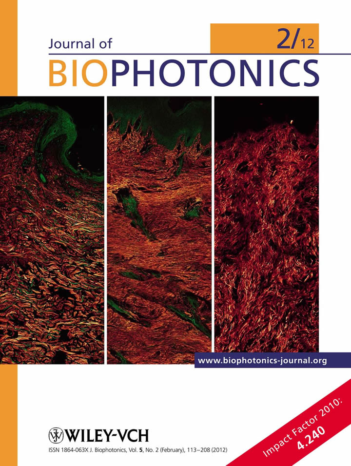
Scar Tissue Classification Using Nonlinear Optical Microscopy and Discriminant Analysis
Scar tissue maturation process occurs stepwise, and calls for reliable classification. The structure of collagen imaged by nonlinear optical microscopy (NLOM) in post-burn hypertrophic, mature scar, and normal skin biopsies, appeared to distinguish these maturation steps. However, it was a discrimination analysis, demonstrated here, that automated and quantified the scar tissue maturation process. The achieved scar classification accuracy was as high as 96%. The combination of NLOM and discrimination analysis is believed to be instrumental in gaining insight into the scar formation, for express diagnosis of scar and surgery planning [1].
1. Kelf TA, Gosnell M, Sandnes B, Guller AE, Shekhter AB, Zvyagin AV: Scar tissue classification using nonlinear optical microscopy and discriminant analysis. Journal of Biophotonics 2012, 5:159-167.
 |
|
Figure. Journal of Biophotonics cover showing three types of collagen architecture in (left to right) normal skin, hypertrophic and mature scar tissue. |
News