All instruments

All instruments

Chromatography

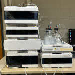
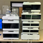
Gel Permeation/Size Exclusion: Agilent 1100 series

Gel Permeation Chromatography (GPC) is a type of Size Exclusion Chromatography (SEC) that separates components of a mixture based on their size.

Agilent 1100 GPC

Used for

Separation and analysis of polymers
Molecular weight and polydispersity of polymers

Specifications

  • Quaternary pump (flow range 0.001-10mL/min)
  • Autosampler (Injection volume 0.1-100 uL, 120 positions sample tray)
  • Diode array and refractive index detectors
  • Mobile phase: THF (HPLC grade provided by user)
  • 2x PLgel 8 um MIXED-C, 7.5 x 300 mm columns
  • Calibrated with polystyrene

Contact

Remi Rouquette
4 Wally’s walk, room 221
Department of Molecular Sciences
Macquarie University
remi.rouquette@mq.edu.au
T: 02 9850 4219

Rates

Discounted rates apply for Macquarie university researchers.

Location

4 Wally’s walk, room 309

Bookings - only after induction and training

HPLC: Agilent 1260 (x2)

The reversed phase HPLC is an instrument used for the separation, identification and quantification of various components in a mixture via UV-vis detection.

Used for

Qualitative analysis
Quantitative analysis
Sample purification and separation

Specifications

  • Quaternary pump (flow range 0.001-10mL/min)
  • Autosampler (Injection volume 0.1-100 uL, 120 positions sample tray)
  • Fraction collector
  • Mobile phase: Water, Acetonitrile or Methanol (HPLC grade provided by user)
  • C18 columns available if needed

Contact

Remi Rouquette
4 Wally’s walk, room 221
Department of Molecular Sciences
Macquarie University
remi.rouquette@mq.edu.au
T: 02 9850 4219

Rates

Discounted rates apply for Macquarie university researchers.

Location

4 Wally’s walk, room 309

Bookings - only after induction and training

Facility homepage

HPLC: Agilent 1290

The reversed phase HPLC is an instrument used for the separation, identification and quantification of various components in a mixture via UV, fluorescence, RID and ELSD detection.

Used for

Qualitative analysis
Quantitative analysis
Sample purification and separation

Specifications

  • Agilent 385 ELSD
  • Fraction Collector analytical scale
  • Infinity Diode-Array Detector
  • Standard Autosampler
  • Infinity Autosampler
  • Infinity Binary Pump
  • Thermostatted Column Compartment
  • Infinity Fluorescence DetectorMobile phase: Water, Acetonitrile or Methanol (HPLC grade provided by user)

Contact

Remi Rouquette
4 Wally’s walk, room 221
Department of Molecular Sciences
Macquarie University
remi.rouquette@mq.edu.au
T: 02 9850 4219

Rates

Discounted rates apply for Macquarie university researchers.

Location

14 Eastern road, room 220 (PC2 lab)

Bookings - only after induction and training

Facility homepage

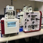
FPLC: Akta Pure

Fast protein liquid chromatography system ideal for the purification and analysis of biomolecules. This system provides high performance, versatility and ease of use for low to medium pressure applications

Used for

Purification and analysis of biomolecules such as proteins using gel filtration, ion-exchange or affinity columns at analytical or preparative scale

Specifications

  • System pump: Piston pump, metering type
  • Flow rate: up to 25 mL/min (0 to 20 MPa operating pressure)
  • UV monitor: 190 - 700nm, up to 3 wavelengths (U9-M)
  • Conductivity monitor
  • Frac9-C fraction collector (Up to 576 fractions, 0.1 to 250 mL fraction volumes)
  • Fraction vessel types:
    • Deep well plates, 96, 48 or 24 wells
    • Tubes 3, 5, 8, 15, 50 ml
    • Bottle, 250 ml

Contact

Remi Rouquette
4 Wally’s walk, room 221
Department of Molecular Sciences
Macquarie University
remi.rouquette@mq.edu.au
T: 02 9850 4219

Location

4 Wally’s walk, room 311 (MUCAB)

Bookings - only after induction and training

Facility homepage

FPLC: Akta Start

The Akta Start is a compact low pressure FPLC system suitable for the separation and analysis of biomolecules. This system provides is highly intuitive and can be operated using a touch screen.

Used for

Quick purification of proteins or buffer exchange using 1-12mL columns eg HiTrap columns

Specifications

  • System pump: Peristaltic pump, single channel
  • Flow rates: up to 5 mL/min (0 - 0.5 MPa operating pressure)
  • Fixed wavelength UV monitor (280 nm)
  • Conductivity monitor
  • Frac30 fraction collector (Up to 30 fractions, 0.5 to 15 mL fraction volumes)
  • Fraction vessel types:
    • 1.5 ml/2 ml microcentrifuge tubes
    • Tubes 5, 12, 15 mL

Contact

Remi Rouquette
4 Wally’s walk, room 221
Department of Molecular Sciences
Macquarie University
remi.rouquette@mq.edu.au
T: 02 9850 4219

Location

4 Wally’s walk, room 311 (MUCAB)

Bookings - only after induction and training

Facility homepage

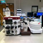
FPLC: Bio-Rad NGC Quest Plus system

The Bio-Rad NGC Quest Plus is a chromatography system for purifying all forms of biomolecules and optimising purification protocols.

Used for

Purification and analysis of biomolecules such as proteins using gel filtration, ion-exchange or affinity columns at analytical or preparative scale

Specifications

  • F10 Pump: Flow rate up to 10 mL/min (0 to 25 MPa operating pressure)
  • Simultaneous multi-wavelength detection of to four wavelengths
  • Conductivity meter
  • BioFrac fraction collector (up to 180 tubes, 0.02 - 250 mL fraction volumes)

Contact

Remi Rouquette
4 Wally’s walk, room 221
Department of Molecular Sciences
Macquarie University
remi.rouquette@mq.edu.au
T: 02 9850 4219

Location

4 Wally’s walk, room 311 (MUCAB)

Bookings - only after induction and training

Facility homepage

GCMS-Shimadzu QP2010

Gas chromatogram mass spectrometer is an instrument used for the separation, identification and quantification of various components in a mixture via flame ionization and mass detection.

Used for

Qualitative analysis
Quantitative analysis
Mass analysis
Only volatile molecules will be detected

Specifications

  • Injection mode: Direct, Autosampler (120 positions sample tray), SPME
  • Single quadrupole (SIM/scan mode, 10 to 1000 m/z, Electron ionization)
  • Flame ionization detector (FID)
  • Mobile phase: UHP Helium (column flow rate 10 mL/min)
  • Large selection of capillary columns available on demand

Contact

Remi Rouquette
4 Wally’s walk, room 221
Department of Molecular Sciences
Macquarie University
remi.rouquette@mq.edu.au
T: 02 9850 4219

Rates

Discounted rates apply for Macquarie university researchers.

Location

4 Wally’s walk, room 202

Bookings - only after induction and training

Facility homepage

GC-MS/MS-Shimadzu TQ8040

Gas chromatogram mass spectrometer is an instrument used for the separation, identification and quantification of various components in a mixture via flame ionization and mass detection.

Used for

Qualitative analysis
Quantitative analysis
Mass analysis
Only volatile molecules will be detected

Specifications

  • Injection mode: Direct, Autosampler (120 positions sample tray), SPME, Headspace
  • Triple quadrupole (SIM, scan, MRM mode, 10 to 1000 m/z, Electron ionization)
  • Flame ionization detector (FID)
  • Mobile phase: UHP Helium (column flow rate 10 mL/min)
  • Large selection of capillary columns available on demand

Contact

Remi Rouquette
4 Wally’s walk, room 221
Department of Molecular Sciences
Macquarie University
remi.rouquette@mq.edu.au
T: 02 9850 4219

Rates

Discounted rates apply for Macquarie university researchers.

Location

4 Wally’s walk, room 202

Bookings - only after induction and training

Facility homepage

LC/MS-Agilent 1260

The reversed phase LC/MS is an instrument used for the separation, identification and quantification of various components in a mixture via UV-vis and mass detection.

Used for

Qualitative analysis
Quantitative analysis
Mass analysis

Specifications

  • Quaternary pump (flow range 0.001-10mL/min)
  • Autosampler (Injection volume 0.1-100 L, 120 positions sample tray)
  • Single quadrupole (SIM/scan mode, positive and negative polarity, ESI-API ionization)
  • Diode Array Detector (190-640 nm)
  • Mobile phase: Water, Acetonitrile or Methanol + 0.1% formic acid (LC/MS grade provided by department)
  • C18 columns available if needed

Contact

Remi Rouquette
4 Wally’s walk, room 221
Department of Molecular Sciences
Macquarie University
remi.rouquette@mq.edu.au
T: 02 9850 4219

Rates

Discounted rates apply for Macquarie university researchers.

Location

4 Wally’s walk, room 311

Bookings

Facility homepage

Coating systems

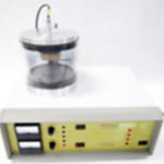
Emitech K550 Gold Sputter Coater

Sputter Coater for preparing specimens for examination by Electron Microscopy

Used for

Ideal for Biological and Material specimens

Specifications

  • Fully automatic control
  • Low voltage Sputtering
  • Cool Sputtering
  • High Resolution Fine Coating (order of 2nm Gold Grain)
  • Special Rotating Stage with full Tilt Facility
  • Even Thickness Deposition (Typically 20nm or 200 Angstroms for SEM work)
  • 165mm Diameter Chamber

Contact

Sue Lindsay
Microscopy Unit Manager
Macquarie University
sue.lindsay@mq.edu.au
T: 02 9850 8124

Rates

Location

14 Eastern Road (E8A), basement level

Facility homepage

Quorum Q 150T ES Carbon/ Chromium Coater

Sputter Coater/Turbo Evaporator for preparing specimens for examination by Electron Microscopy

Used for

Ideal for Biological and Material specimens

Specifications

  • Chromium Sputter coater:
    • Low voltage sputtering
    • Cool sputtering
    • High Resolution Fine Coating (order of 0.5nm Cr grain size)
    • Rotating Stage
    • Even Thickness Deposition (Typically 5nm)
    • 165mm Diameter Chamber
  • Carbon Evaporation:
    • Carbon rod setup
    • Thin films of 5 nm (50A)

Contact

Sue Lindsay
Microscopy Unit Manager
Macquarie University
sue.lindsay@mq.edu.au
T: 02 9850 8124

Rates

Location

14 Eastern Road (E8A), basement level

Facility homepage

Electron Microprobe

Cameca SX 100

Used for

Analysis of multiple elements within polished sections down to the 100 ppm level

Specifications

  • Typical Sample Type: Polished Thin Sections, Disks, or Mounts
  • Tungsten Filament

Contact

Timothy Murphy
Location: E5B - 10 Sir Christopher Ondaatje Avenue, Room 204
Timothy.D.Murphy@mq.edu.au
T: 02 9850 8258

Location

E5B - 10 Sir Christopher Ondaatje Avenue, Room 123

Elemental analyser

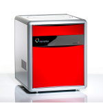
Elementar Cube

Used for

Concentration of Carbon, Nitrogen, and Sulfur in bulk samples

Contact

Olivier Olaard
Location: E5B - 10 Sir Christopher Ondaatje Avenue, Room 203
Olivier.Alard@mq.edu.au

Location

E5B - 10 Sir Christopher Ondaatje Avenue, Room 221

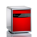
Elementar®- Vario Micro Cube

Analysis of carbon, hydrogen, nitrogen and sulfur content for a wide range of samples.

Used for

C%, H%, N% and S% in sample

Specifications

  • For solid and liquid samples
  • Minimum amount of 2 mg per sample for duplicates.

Contact

Remi Rouquette
4 Wally’s walk, room 221
Department of Molecular Sciences
Macquarie University
remi.rouquette@mq.edu.au
T: 02 9850 4219

Rates

Discounted rates apply for Macquarie university researchers.

Location

10 Sir Christopher Ondaatje Avenue, room 221

Bookings - only after induction and training

Facility homepage

Flow cytometer

BD Influx cell sorter

Flow cytometry is a technique used to detect and measure physical and chemical characteristics of a population of cells or particles based on their optical properties. The BD influx cell sorter include a cell sorting option.

Used for

Cell counting
Cell sorting
Cell characteristics and function
Microorganisms and biomarker detection

Contact

Remi Rouquette
4 Wally’s walk, room 221
Department of Molecular Sciences
Macquarie University
remi.rouquette@mq.edu.au
T: 02 9850 4219

Location

6 Wally’s walk, room 235

Bookings - only after induction and training

Facility homepage

Beckman Coulter CytoFlex S

Flow cytometry is a technique used to detect and measure physical and chemical characteristics of a population of cells or particles based on their optical properties.

Used for

Cell counting
Cell characteristics and function
Microorganisms and biomarker detection

Specifications

  • 96-well plate sample loader
  • Four laser options.

Contact

Remi Rouquette
4 Wally’s walk, room 221
Department of Molecular Sciences
Macquarie University
remi.rouquette@mq.edu.au
T: 02 9850 4219

Rates

Discounted rates apply for Macquarie university researchers.

Location

6 Wally’s walk, room 235

Bookings - only after induction and training

Facility homepage

Microscope

Atomic Force: Bruker Innova AFM

Innova SPM System, Imaging with Contact and Tapping Mode and force spectroscopy, Imaging with derived modes: Lateral Force Microscopy and Phase Imaging
A high performance 5um x 5um closed loop scanner (2.5um Z range)
NanoScope Analysis software, electronics controller and PC Liquid Imaging option

Used for

Accurate, high-resolution and wide range of functionality for advanced research in physical, life and material sciences

Specifications

Contact

Sue Lindsay
Microscopy Unit Manager
Macquarie University
sue.lindsay@mq.edu.au
T: 02 9850 8124

Rates

Location

14 Eastern Road (E8A), basement level

Facility homepage

Brightfield: NIKON Eclipse C1 Microscope

Bright field Microscope

Used for

Ideal for Biological specimens and particle samples

Specifications

  • Ergonomic design
  • LED light
  • CFI Plan Achromat 4x, 10x, 20x, 40x, 100x oil lens
  • CFI Plan Achromat 40x NCG lens
  • DS-Fi3 Colour CMOS USB3.0 camera:
    • high resolution 5.9 megapixels
    • high sensitivity
    • low noise
    • high-speed live display
    • superior colour reproduction

Contact

Sue Lindsay
Microscopy Unit Manager
Macquarie University
sue.lindsay@mq.edu.au
T: 02 9850 8124

Rates

Location

14 Eastern Road (E8A), basement level

Facility homepage

Brightfield: Olympus BX50 Upright Microscope

Bright field Microscope

Used for

Ideal for Biological specimens and material samples

Specifications

  • Routine Bright field microscopy examination
  • Objectives: UPLSAPO10X2 N.A. 0.3,  UPLSAPO20X N.A. 0.75, UPLSAPO40X2 N.A. 0.95, UPLSAPO100X N.A. 1.4
  • CCD 14mp

Contact

Sue Lindsay
Microscopy Unit Manager
Macquarie University
sue.lindsay@mq.edu.au
T: 02 9850 8124

Rates

Location

14 Eastern Road (E8A), basement level

Facility homepage

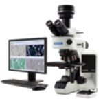
Brightfield: Olympus BX53 Upright Microscope

Bright field Microscope

Used for

Ideal for Biological specimens and particle samples

Specifications

  • Bright field imaging
  • DIC
  • Phase contrast
  • High resolution colour Olympus DP73 digital camera
  • Objectives: UPLFLN4X N.A. 0.13, UPLSAPO10X2 N.A. 0.3,  UPLSAPO20X N.A. 0.75, UPLSAPO40X2 N.A. 0.95, UPLSAPO60X N.A. 1.35, UPLSAPO100X N.A. 1.4, PLCN100X N.A. 1.4 oil.
  • CellSens analytical software

Contact

Sue Lindsay
Microscopy Unit Manager
Macquarie University
sue.lindsay@mq.edu.au
T: 02 9850 8124

Rates

Location

14 Eastern Road (E8A), basement level

Facility homepage

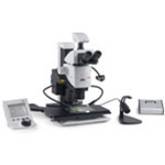
Fluorescence Stereomicroscope: LEICA M205 A

Fluorescence High Resolution  Stereo Microscope

Used for

Ideal for Biological specimens

Specifications

  • 20.5:1 motorized zoom with 7.8x – 320x magnification range and built in display
  • "Fusion Optics” – combines high resolution and depth of field, resulting in high fluorescence sensitivity and ideal 3-D impression
  • Resolution up to 525 lp/mm (1x objective, 10x eyepieces), maximum resolution up to 1050 lp/mm  (2x objective, 10x eyepieces)
  • “Triple Beam” – exclusive fluorescence illumination beam path for homogeneously illuminated, reflex free field of view and excellent signal to noise ratio
  • Fluorescence filter sets: ET DAPI, ET Lucifer Yellow, ET Red and ET mCherry NB
  • Motorized and coded functions: zoom, four position fluorescence filter turret, iris diaphragm and objective nosepiece
  • Fully apochromatic corrected optics
  • Motorized XY Scanning stage
  • Polarisation stage
  • Full LAS X software package
  • 2 Digital Cameras: DFC550 – 12.5 megapixel high resolution CCD, DFC3000 G – 1.3 megapixel Fluorescence Documentation camera
  • LED spotlight illumination, transmitted light and “Flexi Dome” Diffuse illumination

Contact

Sue Lindsay
Microscopy Unit Manager
Macquarie University
sue.lindsay@mq.edu.au
T: 02 9850 8124

Rates

Location

14 Eastern Road (E8A), basement level

Facility homepage

Fluorescence Inverted Confocal: Olympus FluoView FV 1000 IX81

Laser Scanning Confocal Microscope

Used for

Ideal for Biological specimens and biological material samples

Specifications

  • Versatile point-scanning, point-detection, confocal laser scanning microscope
  • Ideal for 3-D imaging, time course experiments, energy transfer visualisation and photobleaching recovery experiments
  • Delivers the High resolution required for multi-dimensional observation of cell and tissue morphology as well as precise molecular localisation
  • Real time visualisation of rapid cell responses
  • Able to measure diffusion coefficients of intracellular molecules and quantifying molecular kinetics
  • 4 laser diodes – 405, 473, 559 and 635nm
  • DIC
  • UIS 2 objectives: UPLSAPO10x2 N.A. 0.4, UPLSAPO20X N.A. 0.75, UPLSAPO40X2 N.A. 0.95, UPLSAPO 60X N.A. 1.35 oil, PLAPON 60xO N.A 1.42BF oil
  • 3 fluorescence channels, 1 transmitted light channel, sequential multi-channel acquisition of up to 10 channels
  • Time lapse imaging
  • The environmental chamber enables temperature, humidity and carbon dioxide regulation, allowing long time-course experiments with live cell

Contact

Sue Lindsay
Microscopy Unit Manager
Macquarie University
sue.lindsay@mq.edu.au
T: 02 9850 8124

Rates

Location

14 Eastern Road (E8A), basement level

Facility homepage

Fluorescence Inverted Confocal: Olympus FluoView FV 3000RS IX83

High Resolution Live Imaging Laser Scanning Confocal Microscope

Used for

Ideal for Biological specimens and nanobiological/particle samples

Specifications

  • Imaging diverse fluorophores in fixed or live samples
  • Temperature, humidity, CO2 control
  • Spectrally tunable detectors
  • Ultra-fast resonant scanning (reduced photo-damage with live samples)
  • 3D z-stacks, timelapse, multi-position timelapse
  • Olympus IX83 fully motorized inverted microscope
  • All Diode Lasers with 4 Laser Lines–405 nm, 488 nm, 561 nm and 640 nm

Contact

Sue Lindsay
Microscopy Unit Manager
Macquarie University
sue.lindsay@mq.edu.au
T: 02 9850 8124

Rates

Location

14 Eastern Road (E8A), basement level

Facility homepage

Fluorescence: Olympus BX 63

Fluorescence Upright Microscope

Used for

Ideal for Biological specimens and particle samples

Specifications

  • Fully motorised system features a programmable ultrasonic scanning stage allowing for automated multidimensional and multichannel imaging
  • Highly sensitive dual colour and monochrome digital camera, ideal for fluorescence and brightfield applications
  • Fluorescence; wide band UV (BP330-385), Wide Band Blue (BP450-480), Wide Band Interference Green (BP530-550)
  • Objectives: UPLSAPO4X N.A. 0.16, UPLSAPO10X2 N.A. 0.40, UPLSAPO20X N.A. 0.75, UPLSAPO40X N.A. 0.95, UPLFLN60X N.A. 0.9, UPLSAPO100X N.A. 1.4 oil
  • DIC
  • Brightfield
  • CellSens image analysis software

Contact

Sue Lindsay
Microscopy Unit Manager
Macquarie University
sue.lindsay@mq.edu.au
T: 02 9850 8124

Rates

Location

14 Eastern Road (E8A), basement level

Facility homepage

Macro and micro high resolution imaging system: BK PLUS Lab System

Macro & Micro High Resolution Imaging System

Used for

Ideal for Biological, Geological, Archaeological and Material samples

Specifications

  • A Macro and Micro Imaging System
  • Imaging Specimen size ranging from 60cm to 60µm
  • This microscope lens will allow you to image clear measurable detail in brilliant colour down to 25 microns
  • Visionary Digital P-51Cam-Lift ultra high precision Linear Actuator
    P-51Cam-Lift camera stand-off with quick release bracket
    Visionary Digital Linear actuator software with image composite settings
    Linear actuator controller with high resolution micro stepper and adjustable optical limit switches. (Resolves to less than 0.5 micron)
  • Canon 5DS (50 Megapixels) Macro camera
  • Canon 7D Mark II (20 Megapixels) Micro camera
  • Microscope lens:  
    5X LD Mitutoyo objective
    10X LD Mitutoyo objective
    20X LD Mitutoyo objective
  • Canon 35mm Wide angle professional lens
  • Canon f2.5 50mm Macro-photography lens
  • Customized Canon MPE 65mm 1-5X micro-photography lens
  • Customized Canon EF 100mm f2.8 IS USM Professional macro lens
  • 1.4X Teleconverter
  • 1600 Watt Power pack for total control of Pro Studio flash heads
  • Flash sync speeds to 1/8000 sec
  • Professional studio flash heads with Modeling Lights
  • Software:
    Our Passport II software
    Phase One Capture One imaging software with 3 seats.
    Photoshop CS6Extended with all loaded pre-sets and measurement
    Zerene Stacker
    Canon EOS Utilities
  • The patented  FX3 Fiber Optic Flash Illumination System

Contact

Sue Lindsay
Microscopy Unit Manager
Macquarie University
sue.lindsay@mq.edu.au
T: 02 9850 8124

Rates

Location

14 Eastern Road (E8A), basement level

Facility homepage

Scanning electron microscope: Field Emission SEM: Jeol JSM 7100F FESEM

The Jeol JSM 7100F FESEM has a resolution (sample dependent) of 1.2nm at 30kV and 3.0nm at 1kV

Used for

Ideal for imaging of micro and nano structures, both biological and material samples.

Specifications

  • Equipped with a Kleindiek MM3A-EM Micromanipulator for precision in-situ nanomanipulation
  • Equipped with EX-94300 SDD Energy Dispersive X-ray analyser

Contact

Sue Lindsay
Microscopy Unit Manager
Macquarie University
sue.lindsay@mq.edu.au
T: 02 9850 8124

Rates

Location

14 Eastern Road (E8A), basement level

Facility homepage

Scanning electron microscope: FEI Field Emission SEM with Nanomin software

Used for

High-resolution imaging and mineral/EDX mapping

Specifications

  • Typical Sample Type: Polished Thin Sections, Disks, or Mounts
  • Field Emission
  • 5-30 kV Emission
  • Secondary Electron and Backscatter imaging
  • 2x Bruker XFlash 6|30 EDX Detectors
  • Liquid Nitrogen Condenser

Contact

Sean Murray
E5A - 11 Wally's Walk, Room B11 (MUSSIL)
Sean.Murray@mq.edu.au
T: 02 9850 6121

Location

E5A – 11 Wally’s Walk, Room B11

Scanning electron microscope: Tungsten Variable Pressure SEM: JEOL 6480LA

Variable pressure SEM (LV pressure -1 to 270 Pa). Maximum resolution of 4 nm, with a large depth of field. Suitable for analysis of biological and material specimens.

Used for

Great for uncoated biological specimens, also suitable for material specimens.

Specifications

  • Equipped with a Deben Coolstage

Contact

Sue Lindsay
Microscopy Unit Manager
Macquarie University
sue.lindsay@mq.edu.au
T: 02 9850 8124

Rates

Location

14 Eastern Road (E8A), basement level

Facility homepage

Scanning electron microscope: benchtop SEM: Phenom XL

Easy to use multi-functional desktop Scanning Electron Microscope.

Used for

For high-throughput imaging and elemental analysis of biological and material samples, devices and components.

Specifications

  • Electron magnification range: 80 – 100,000x
  • Resolution: ≤ 20nm
  • Acceleration voltages: 5kV, 10kV and 15kV
  • Vacuum modes: standard, charge reduction and high vacuum
  • Detectors: BSD (backscattered), SED (secondary) and EDS
  • Sample size: max. 100mm x 100mm. Up to 36 x 12mm pin stubs
  • EDS mapping, spot and line analysis
  • Real time in-situ tensile testing (MICROTEST module)
  • In-situ chemical probing/molecular imaging of a sample in liquid (Wet Cell II module)

Contact

Sue Lindsay
Microscopy Unit Manager
Macquarie University
sue.lindsay@mq.edu.au
T: 02 9850 8124

Rates

Location

14 Eastern Road (E8A), basement level

Facility homepage

Scanning Electron Microscope: benchtop SEM: Jeol NEOscope 6000plus

Easy to use, moveable benchtop Scanning Electron Microscope. High and low vacuum modes – non-destructive in low vacuum mode, and no preparation needed for samples in low vacuum mode.

Used for

For portable imaging of biological and material samples, devices and components.

Specifications

  • Electron magnification range: 10 – 60,000x
  • Acceleration voltages: 5kV, 10kV and 15kV
  • Vacuum modes: High and Low vacuum modes
  • Detectors: BSD (backscattered), SED (secondary) and EDS
  • EDS mapping, spot and line analysis.
  • Non-destructive specimen analysis in low-vacuum mode
  • NO preparation to specimens in low-vacuum mode needed
  • Ability to start in low-vacuum mode
  • Manual specimen stage
  • Tilt/rotation motor drive specimen chamber
  • Sample size: max. 70mm x 50mm.
  • Full- auto, filament adjustment, alignment, focus, stigmator, exposure

Contact

Sue Lindsay
Microscopy Unit Manager
Macquarie University
sue.lindsay@mq.edu.au
T: 02 9850 8124

Rates

Location

14 Eastern Road (E8A), basement level

Facility homepage

Scanning electron microscope: Zeiss EVO|MA15 SEM with Aztec Software

Used for

High-resolution imaging with a specialization in elemental mapping and EBSD

Specifications

  • Typical Sample Type: Polished Thin Sections, Disks, or Mounts
  • Tungsten Filament
  • Backscatter and Secondary Electron Imaging
  • Oxford Instruments X-Max EDX
  • Horiba iHR320 EBSD

Contact

Timothy Murphy
Location: E5B - 10 Sir Christopher Ondaatje Avenue, Room 204
Timothy.D.Murphy@mq.edu.au
T: 02 9850 8258

Location

E5B - 10 Sir Christopher Ondaatje Avenue, Room 123

Shape Analyser: Kruss Drop DSA30

The Drop Shape Analyser DSA30 measuring system

Used for

  • Automatic control of the instrument and data recording by user-defined test methods
  • Measurements at low and high temperatures
  • Evaluation of a digitised drop image by various methods
  • Calculation of the surface energy of a solid by various methods
  • Integrated and extendable databases with substance data on liquids that can be directly linked to the measuring data
  • Comfortable data management and documentation

Contact

Sue Lindsay
Microscopy Unit Manager
Macquarie University
sue.lindsay@mq.edu.au
T: 02 9850 8124

Rates

Location

14 Eastern Road (E8A), basement level

Facility homepage

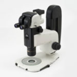
Stereomicroscope: NIKON SMZ18

Stereo Microscope

Used for

Ideal for Biological, Geological, Archaeological and Material samples

Specifications

  • Manual Zoom
  • Ergonomic design and trinocular tilting head
  • Observation: Brightfield and simple polarizing
  • Zoom ratio: 18:1
  • Magnification range: 0.75x – 13.5x
  • P-Plan 1x (W.D. 78mm) objective lens
  • Manual XY stage
  • DS-Fi3 Colour CMOS USB3.0 camera:
    • high resolution 5.9 megapixels
    • high sensitivity
    • low noise
    • high-speed live display
    • superior colour reproduction

Contact

Sue Lindsay
Microscopy Unit Manager
Macquarie University
sue.lindsay@mq.edu.au
T: 02 9850 8124

Rates

Location

14 Eastern Road (E8A), basement level

Facility homepage

Stereomicroscope: Olympus SZX16

Stereo Microscope

Used for

Ideal for Biological, Geological, Archaeological and Material samples

Specifications

  • Research Stereo Microscope
  • Zoom 16.4:1
  • Objectives 1x and 2x
  • Transmitted, collar and gooseneck LED illumination
  • Digital colour camera Olympus DP26
  • CellSens imaging software

Contact

Sue Lindsay
Microscopy Unit Manager
Macquarie University
sue.lindsay@mq.edu.au
T: 02 9850 8124

Rates

Location

14 Eastern Road (E8A), basement level

Facility homepage

Transmission Electron Microscope: Philips CM10 TEM

Transmission electron microscope, with a resolution of up to 0.2 nm

Used for

Ideal for Biological specimens and nanoparticle samples

Specifications

  • 100 KV with resolution of up to 0.2 nm
  • Olympus SIS Megaview G2 digital camera has a resolution of 1376 x 1032 pixels with a 14-bit dynamic range (16384 grey values)
  • Measurement and analysis functions are available in Olympus Radius software

Contact

Sue Lindsay
Microscopy Unit Manager
Macquarie University
sue.lindsay@mq.edu.au
T: 02 9850 8124

Rates

Location

14 Eastern Road (E8A), basement level

Facility homepage

Zoom microscope: Microeye Discovery Micros

Educational Zoom Microscope

Used for

Ideal for Biological, Geological, Archaeological and Material samples

Specifications

  • Educational microscopes for school groups
  • Extremely robust and hardwearing
  • LED lighting
  • Ease of use
  • Magnification of 2x to 40x
  • Examine of differing heights (up to 100mm)

Contact

Sue Lindsay
Microscopy Unit Manager
Macquarie University
sue.lindsay@mq.edu.au
T: 02 9850 8124

Rates

Location

14 Eastern Road (E8A), basement level

Facility homepage

Specimen preparation instruments

Leica EM CPD 300 Critical Point Dryer

Critical Point Dryer

Used for

Specimen preparation for SEM examination

Specifications

  • Fully automatic critical point dryer
  • Used to dry biological and micro-electro-mechanical samples without distortion
  • Fully reproducible processes through full automation
  • Highly reproducible sample preparation
  • Reduced process times
  • Minimal user interaction time through automation and intuitive software
  • Ease of use y intuitive software and integrated touch screen
  • Store and recall drying programs
  • Flexibility in sample size with large variety of sample holders

Contact

Sue Lindsay
Microscopy Unit Manager
Macquarie University
sue.lindsay@mq.edu.au
T: 02 9850 8124

Rates

Location

14 Eastern Road (E8A), basement level

Facility homepage

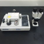
Gatan Dimple Grinder model 656

Material specimen preparation for TEM examination

Used for

Material specimen preparation for TEM examination

Specifications

  • Precision instrument used to produce circular dimples (spherical or flat-bottomed profile) in materials such as ceramics, semiconductors, metals and combinations thereof.
  • *Other applications include the production of accurate, precisely-located taper sections through the surface of specimens, which may then be subjected to analytical techniques such as EDX, SIMS, Auger etc. In this way compositional profiles of segregated surface layers, coating and plantings can be obtained rapidly and with excellent “depth resolution.”

Contact

Sue Lindsay
Microscopy Unit Manager
Macquarie University
sue.lindsay@mq.edu.au
T: 02 9850 8124

Rates

Location

14 Eastern Road (E8A), basement level

Bookings

Facility homepage

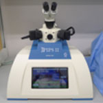
Gatan Pips II Model 695 Precision Ion Polishing System

Precision ion polishing system for precise centering, control and reproducibility of your milling process.

Used for

Ideal for Material specimens

Specifications

  • Compact, benchtop system designed to produce high-quality TEM specimens with exceptionally large, clean, electron-transparent areas.
  • X,Y stage permits alignment of argon beams to region on the sample
  • Improved collimated beam provides useable voltages as low as 100 volts for rapid and damage free preparation of FIB lamella
  • 10” colour touch screen for display and control of all PIPS II parameters

Contact

Sue Lindsay
Microscopy Unit Manager
Macquarie University
sue.lindsay@mq.edu.au
T: 02 9850 8124

Rates

Location

14 Eastern Road (E8A), basement level

Facility homepage

Leica EM UC7 Ultramicrotome with CCD

Ultramicrotome

Used for

Ideal for Semi-thin and Ultra-thin sections for SEM, TEM and LM examination

Specifications

  • Ergonomic design
  • Fully configurable systems
  • Motorised knife stage
  • Stereo microscope 8:1 zoom
  • IC80 HD digital camera for live viewing

Contact

Sue Lindsay
Microscopy Unit Manager
Macquarie University
sue.lindsay@mq.edu.au
T: 02 9850 8124

Rates

Location

14 Eastern Road (E8A), basement level

Facility homepage

Reichert Ultracut S microtome

Ultramicrotome

Used for

Ideal for Semi-thin and Ultra-thin sections for SEM, TEM and LM examination

Specifications

  • Ultra-thin sections (80nm) for Transmission electron microscopy
  • Three trans illumination pathways (top, under and behind specimen) facilitate viewing of the specimen
  • Stereo zoom microscope, magnification 10x to 60x

Contact

Sue Lindsay
Microscopy Unit Manager
Macquarie University
sue.lindsay@mq.edu.au
T: 02 9850 8124

Rates

Location

14 Eastern Road (E8A), basement level

Facility homepage

Leica VT 1000S Vibratome

Vibramicrotome

Used for

Ideal for Fresh or fixed, animal or plant tissues

Specifications

  • A vibrating blade microtome
  • Sections fresh or fixed, animal or plant tissues
  • Can produce Fresh tissue sections in order of 20um that permit enhanced optical resolution
  • The combined use of Vibratome sections and the Inverted Confocal Microscope system allows for imaging experiments to be performed with the high numerical aperture immersion objectives
  • The Vibratome facilitates experimental work where cellular visualisation is critical

Contact

Sue Lindsay
Microscopy Unit Manager
Macquarie University
sue.lindsay@mq.edu.au
T: 02 9850 8124

Rates

Location

14 Eastern Road (E8A), basement level

Facility homepage

Knifemarker LKB 2178

Glass knife maker for use on the Ultramicrotomes

Used for

Glass knife maker for use on the Ultramicrotomes

Contact

Sue Lindsay
Microscopy Unit Manager
Macquarie University
sue.lindsay@mq.edu.au
T: 02 9850 8124

Rates

Location

14 Eastern Road (E8A), basement level

Facility homepage

Reichert Jung 1140/Autocut microtome

Universal automatic and manual microtome

Used for

Ideal for Biological and Material specimens

Specifications

  • Universal automatic and manual microtome
  • Stereo microscope
  • Sectioning resin embedded and paraffin-embedded materials
  • Section thickness is adjustable from 3µm to 35µm
  • Glass knives, disposable steel knives and tungsten carbide knives

Contact

Sue Lindsay
Microscopy Unit Manager
Macquarie University
sue.lindsay@mq.edu.au
T: 02 9850 8124

Rates

Location

14 Eastern Road (E8A), basement level

Facility homepage

Reichert Om E Large Sliding Microtome

Sectioning of hard or large materials, such  as wood, stems, bone, plastics and large muscles

Used for

Ideal for Sectioning of hard or large materials, such  as wood, stems, bone, plastics and large muscles

Specifications

  • Sectioning of hard or large materials, such  as wood, stems, bone, plastics and large muscles
  • Section thickness 1µ to 30µ
  • Specimen blocks up to 35 x 45mm can be held

Contact

Sue Lindsay
Microscopy Unit Manager
Macquarie University
sue.lindsay@mq.edu.au
T: 02 9850 8124

Rates

Location

14 Eastern Road (E8A), basement level

Facility homepage

American Optical Spencer Rotary Microtome 820

Sectioning of paraffin embedded material for histology

Used for

Ideal for Biological specimens

Specifications

  • Sectioning of paraffin embedded material for histology
  • Manual use
  • Specimens up to 32mm by 27mm
  • Section thickness is adjustable from 1µm to 50µm

Contact

Sue Lindsay
Microscopy Unit Manager
Macquarie University
sue.lindsay@mq.edu.au
T: 02 9850 8124

Rates

Location

14 Eastern Road (E8A), basement level

Facility homepage

Leica EG1150 Modular Tissue Embedding station

Paraffin wax embedding station

Used for

Ideal for Biological specimens

Specifications

  • Paraffin wax embedding station
  • Cold plate to allow manipulation of the specimen to give correct cutting orientation
  • Multiple cassette system

Contact

Sue Lindsay
Microscopy Unit Manager
Macquarie University
sue.lindsay@mq.edu.au
T: 02 9850 8124

Rates

Location

14 Eastern Road (E8A), basement level

Facility homepage

Selfrag

Used for

Rock disaggregation using high-voltage electrical pulses

Specifications

  • Typical Sample Type: 1 kg of crushed rock, but can do significantly less material

Contact

Sean Murray
10 Sir Christopher Ondaatje Ave, Room 216
sean.murray@mq.edu.au
T: 02 9850 4729

Location

E5A - 11 Wally's Walk, Room B31

Spectrometer

Circular dichroism: Jasco J-1500

The J-1500 circular dichroism (CD) spectropolarimeter allows simultaneous measurement of 4 modes including CD, linear dichroism (LD), and absorbance. We have a range of accessories including long pass cut-off filters for excitation fluorescence spectroscopy, film stretchers for LD, Couette flow cells for LD, multiple -position thermostatted cuvette holder. Users are generally expected to provide their own quartz cuvettes

Circular dichroism (CD) measures the difference in absorbance of left- and right- handed polarized light of chiral molecules, which is commonly used to analyse the conformation of macromolecules, particularly protein secondary structure.
Linear dichroism (LD) measures the difference in absorption of light linearly polarised parallel and perpendicular to the orientation axis. LD can be used to give information about the conformation and orientation of structures with orientated molecules such as DNA complexes, fibrous proteins such as (e.g. cytoskeleton and amyloid proteins) and membrane peptides/proteins in liposomes.
The J-1500 is a hybrid instrument consisting of a variable wavelength polarimeter for CD or LD and absorption spectrophotometer (~180-900 nm, Far-UV to Vis range). The J-1500 is fitted with a 6-position multi-cell changer and Peltier temperature control unit. This set-up is ideally suited to:

  • Determine protein secondary structure and analyse conformational changes
  • Nucleic acid structure determination and changes upon binding or melting
  • Other thermodynamic parameters and kinetics based measurements

Used for

Routine solution state experiments on chiral molecules with a range of more sophisticated experiments possible including temperature controlled, kinetics, linear dichroism, fluorescence detected CD and LD.

Specifications

  • Operates from 950 – 163 nm
  • Light source : 150 W air-cooled Xenon lamp or 450 W water-cooled Xenon lamp (factory-installed option)
  • Detector : Head-on photomultiplier tube PMT
  • Optional photomultiplier tube PMT
  • Optional InGaAs
  • Modulator : Piezoelastic modulator
  • Measurement wavelength range : 163 to 950 nm
  • Wavelength accuracy : 0.1 nm from 163 to 250 nm
  • 0.2 nm from 250 to 500 nm
  • 0.5 nm from 500 to 800 nm
  • Spectral bandwidth: 0.01 to 16 nm
  • Slit width : 1 to 4000 μm
  • Digital Integration Time (D.I.T.) : 0.1 msec to 30 sec.
  • Scanning mode : Continuous scan
    • Step scan
    • Auto response(D.I.T) scan
  • Scanning speed : up to 10000 nm/min

Contact

Alison Rodger or Sophie Goodchild
4 Wally’s walk, laboratory 302
Department of Molecular Sciences
Macquarie University
sophie.goodchild@mq.edu.au
T: 02 9850 8235

Rates

Discounted rates apply for Macquarie university researchers.

Location

4 Wally’s walk, room 302 (restricted access)

Bookings

Facility homepage

Circular dichroism: Jasco J-810

Circular dichroism (CD) measures the difference in absorbance of left- and right-handed polarized light, which can be a powerful tool for identification and characterisation of chiral molecules. The J-810 is a hybrid instrument consisting of a variable wavelength polarimeter and absorption spectrophotometer (~180-900 nm, Far-UV to Vis range). Our J-810 is also fitted with an Optical Rotation Dispersion (ORD) module and Peltier temperature control unit making it ideally suited to measure optical purity/identification of small molecules and natural products chemistry.

Specifications

  • Light source: 150 W Xenon arc lamp (air cooled)
  • Wavelength range: ~180 to ~900 nm (standard)
  • Four Channel Simultaneous Data Acquisition: Input channels: 2 internal plus 2 external signals can be acquired simultaneously
  • ORDM-401 unit fitted

Contact

Alison Rodger or Sophie Goodchild
4 Wally’s walk, laboratory 302
Department of Molecular Sciences
Macquarie University
sophie.goodchild@mq.edu.au
T: 02 9850 8235

Rates

Discounted rates apply for Macquarie university researchers.

Location

4 Wally’s walk, room 302 (restricted access)

Bookings

Facility homepage

Infra-red: Jasco FT/IR-4700

The FT/IR 4700 is a Mid-IR Fourier Transform Infrared (FTIR) spectrometer (wavenumber range 7800 to 350 cm-1) with high resolution (0.4cm-1 and S/N over 35,000:1), making it ideal for transmission measurements of solid, liquid and gas samples. A real-time data processing function can be used to observe and verify the spectrum during measurements. The FT/IR 4700 can also be paired with the PIKE MIRacle™ Single Reflection ATR accessory with ZeSe crystal for high throughput attenuated total reflectance (ATR) measurements.

Specifications

  • Light source: High-intensity ceramic source
  • Wavenumber Range: 7,800 to 350 cm-1
  • Wavenumber accuracy: ± 0.01 cm-1 (theoretical value)
  • Maximum resolution: 0.4 cm-1
  • Optical System: Single beam
  • Sample chamber: Size: 200 mm (W) × 260 mm (D) × 185 mm (h) (can be nitrogen purged)

Contact

Alison Rodger or Sophie Goodchild
4 Wally’s walk, laboratory 302
Department of Molecular Sciences
Macquarie University
sophie.goodchild@mq.edu.au
T: 02 9850 8235

Rates

Location

4 Wally’s walk, room 302 (restricted access)

Bookings

Facility homepage

Light Scattering: Malvern Zeta Sizer Nano SP

The Malvern Zetasizer Nano is a light scattering system suitable for characterisation of colloid dispersions, nanoparticles and macromolecules such as polymer and protein solutions (3.8 nm to 100 μm, concentration ~0.1ppm to 40%w/v, pH 1-13, sample dependent).  Three different light scattering measurements can be performed:

  • Dynamic Light Scattering (DLS) – is used to measure the diffusion of particles under Brownian motion, and convert this to a size distribution using the Stokes-Einstein relationship. Non-invasive back-scatter (NIBS) technology is incorporated to give better sensitivity and the ability to measure particles at high concentration.
  • Electrophoretic Light scattering (ELS) – is used to measure zeta potential. An electric field is applied to the sample and mixed mode measurement, phase analysis light scattering (M3-PALS) is used to measure electrophoretic mobility and determine a zeta potential distribution.
  • Static Light Scattering (SLS) – is used to determine the molecular weight (> ~1KDa) of proteins and polymers. The scattering intensity at a range of concentrations is measured to construct a Debye plot. From this the weight-average molecular weight and second virial coefficient can be calculated to provide a measure of protein solubility.

Specifications

  • Measurement range: 3.8nm – 100 microns diameter (sample dependent)
  • Measurement principle: Electrophoretic Light Scattering
  • Minimum sample volume: 150µL (20µL using diffusion barrier method)
  • Accuracy: 0.12µm.cm/V.s for aqueous systems using NIST SRM1980 standard reference material
  • Sensitivity: ~0.1-10mg/mL (BSA/Lysozyme)
  • Temperature control range: 0°C - 90°C +/-0.1
  • Light source: He-Ne laser 633nm, Max 4mW

Contact

Alison Rodger or Sophie Goodchild
4 Wally’s walk, laboratory 302
Department of Molecular Sciences
Macquarie University
sophie.goodchild@mq.edu.au
T: 02 9850 8235

Rates

Discounted rates apply for Macquarie university researchers.

Location

4 Wally’s walk, room 302 (restricted access)

Bookings

Facility homepage

Mass spectrometer: ICP-MS: Agilent 7500 Series

Used for

ppb level resolution on trace elements in acidified solutions

Specifications

  • Typical Sample Type: Dissolved Material in 2% HCl or HNO3
  • Integrated Autosampler
  • Solution Only

Contact

Peter Wieland
E5B - 10 Sir Christopher Ondaatje Avenue, Room 206
Peter.Wieland@mq.edu.au
T: 02 9850 4402

Location

E5B - 10 Sir Christopher Ondaatje Avenue, Room 126

Mass spectrometer: ICP-MS: Agilent 7700 Series

Used for

High-throughput ICP-MS

Contact

Yi-Jen Lai
E5B - 10 Sir Christopher Ondaatje Avenue, Room 204
Yi-Jen.Lai@mq.edu.au
T: 02 9850 8376

Locations

E5B - 10 Sir Christopher Ondaatje Avenue, Room 106
E5B - 10 Sir Christopher Ondaatje Avenue, Room 126

Mass spectrometer: ICP-MS: Agilent 8900 Triple Quad

Used for

Trace elements in studies where isobaric interferences are a concern

Contact

Yi-Jen Lai
E5B - 10 Sir Christopher Ondaatje Avenue, Room 204
Yi-Jen.Lai@mq.edu.au
T: 02 9850 8376

Location

E5B - 10 Sir Christopher Ondaatje Avenue, Room 108

Mass spectrometer: ICP-MS: Nu Attom High-Resolution ICP-MS

Used for

High-resolution isotope-ratios in a solid matrix using laser ablation

Specifications

  • Typical Sample Type: Polished thin sections or disks
  • Attached to a Photon-Machines Laser Ablation System for Spot Analysis
  • Laser Ablation Only

Contact

Yi-Jen Lai
E5B - 10 Sir Christopher Ondaatje Avenue, Room 204
Yi-Jen.Lai@mq.edu.au
T: 02 9850 8376

Location

E5B - 10 Sir Christopher Ondaatje Avenue, Room 126

Mass spectrometer: ICP-MS: Nu Plasma II

Used for

Multicollector ICP-MS

Contact

Yi-Jen Lai
E5B - 10 Sir Christopher Ondaatje Avenue, Room 204
Yi-Jen.Lai@mq.edu.au
T: 02 9850 8376

Location

E5B - 10 Sir Christopher Ondaatje Avenue, Room 106

Mass spectrometer: ICP-MS: Nu Plasma HR

Used for

High Resolution Multi-Collector ICP-MS

Contact

Yi-Jen Lai
E5B - 10 Sir Christopher Ondaatje Avenue, Room 204
Yi-Jen.Lai@mq.edu.au
T: 02 9850 8376

Location

E5B - 10 Sir Christopher Ondaatje Avenue, Room 108

Mass spectrometer: ICP-MS: Thermo Fisher Neptune High Resolution ICP-MS

Used for

High-Resolution ICP-MS

Specifications

  • 10x13 Ω resistors

Contact

Peter Wieland
E5B - 10 Sir Christopher Ondaatje Avenue, Room 206 
Peter.Wieland@mq.edu.au
T: 02 9850 4402

Location

E5B - 10 Sir Christopher Ondaatje Avenue, Room 135

Mass spectrometer: Isotope ratio MS MAT 253 plus with IBEX

Used for

MAT 253 plus MS: δ13C, δ18O, and Δ47 measurements on CO2 gas IBEX: Acid digestion and purification of carbonates for direct input into MAT 253 plus MS

Specifications

  • Typical Sample Type: 2-4 mg pure carbonate aliquots
  • 7 Faraday cup configuration
  • 1013Ω resistors on the mass 47, 47.5, 48, and 49 cups
  • Dual-Inlet
  • IBEX sample preparation system with controlled temperature reactions in a common acid bath

Contact

Sean Murray
10 Sir Christopher Ondaatje Ave, Room 216
sean.murray@mq.edu.au
T: 02 9850 4729

Location

E5A – 11 Wally’s Walk, Room B11

Mass spectrometer: Thermal ionization MS: Thermo Triton

Used for

Measuring isotopic ratios that are used in geochronology, cosmochemistry, and geochemistry. e.g. Sr-Nd, U-Th-Pb, U-series disequilibrium, Sr, Nd, Hf, & Os in seawater

Specifications

  • Typical Sample Type: Powdered bulk material that is acid digested and chemically cleaned

Contact

Peter Wieland
E5B - 10 Sir Christopher Ondaatje Avenue, Room 206
Peter.Wieland@mq.edu.au
T: 02 9850 4402

Location

E5B - 10 Sir Christopher Ondaatje Avenue, Room 124

Mass spectrometer: Thermal ionization MS: Thermo Fisher Triton Plus

Used for

High Precision Isotope Ratios

Contact

Peter Wieland
E5B - 10 Sir Christopher Ondaatje Avenue, Room 206 
Peter.Wieland@mq.edu.au
T: 02 9850 4402

Location

E5B - 10 Sir Christopher Ondaatje Avenue, Room 135

MP-AES – Agilent 4100

Microwave induced Plasma Atomic Emission Spectrophotometer (MP-AES) measures levels of  elements in solution.

Used for

Simultaneous multi-analyte determination of major and minor elements. Working range of low ppm to weight percent (wt. %).

Specifications

  • Nitrogen supplied
  • Samples must be digested in low concentrated nitric acid
  • A wide range of ICP grade standards available in teaching lab
  • Similar to ICP-MS, AAS.

Contact

Remi Rouquette
4 Wally’s walk, room 221
Department of Molecular Sciences
Macquarie University
remi.rouquette@mq.edu.au
T: 02 9850 4219

Rates

Discounted rates apply for Macquarie university researchers.

Location

14 Sir Christopher Ondaatje Avenue, room 321.

Bookings please contact Dr Remi Rouquette

Facility homepage

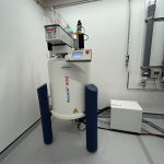
Nuclear Magnetic Resonance: Bruker AVIIIHD 400 MHz NMR Spectrometer

The Avance 400 MHz NMR spectrometer, with the SampleXpress 60 Holder Autosampler and Iconnmr automation software, is the workhorse of the NMR facility. Samples are conveniently submitted and queued for analysis.

Bruker AVIIIHD 400 MHz NMR Spectrometer

Used for

Routine solution state 1D and 2D experiments on small molecules

Specifications

  • 5mm BBFO SmartProbe™ (19F/31P-109Ag)
  • Actively-shielded high performance Z-gradient
  • 19F{1H} and 2D 1H/19F correlation spectroscopy
  • Operating temperature range: -150°C to +150°C
  • Ascend 400 MHz (9.4 T) Magnet
  • Drift rate < 4.0 Hz/hr
  • BCUII (Bruker Chiller Unit)
  • Accessible temperatures ≥ 200 K
  • SampleXpress 60 Holder Autosampler

Contact

Remi Rouquette
4 Wally’s walk, room 221
Department of Molecular Sciences
Macquarie University
remi.rouquette@mq.edu.au
T: 02 9850 4219

Rates

Discounted rates apply for Macquarie university researchers.

Location

4 Wally’s walk, room 014 (highly restricted access)

Bookings

Facility homepage

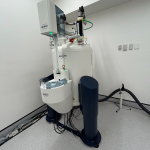
Nuclear Magnetic Resonance: Bruker AVIIIHD 500 MHz NMR Spectrometer

The Avance 500 MHz NMR spectrometer is set up to run variable temperature experiments (-70°C to 100°C) routinely. Very low temperature experiments (-70°C to -150°C) can also be run with use of the liquid nitrogen evaporator and staff assistance. It is highly sensitive for carbon detection, thus it is often utilized to acquire data for the complete characterization of small to medium sized organic molecules.

Bruker AVIIIHD 500 MHz NMR Spectrometer

Used for

Variable temperature solution state experiments, characterization experiments

Specifications

  • 5mm BBFO SmartProbe™ (19F/31P-109Ag)
  • Actively-shielded high performance Z-gradient
  • 19F{1H} and 2D 1H/19F correlation spectroscopy
  • Operating temperature range: -150°C to +150°C
  • Ascend 500 MHz (11.7 T) Magnet
  • Drift rate < 5.0 Hz/hr
  • Magnet leg upgrade to damp vibrations above 3.8 Hz
  • BCUII (Bruker Chiller Unit)
  • Accessible temperatures ≥ 200 K
  • SampleCase 24 Holder Autosampler

Contact

Remi Rouquette
4 Wally’s walk, room 221
Department of Molecular Sciences
Macquarie University
remi.rouquette@mq.edu.au
T: 02 9850 4219

Rates

Discounted rates apply for Macquarie university researchers.

Location

4 Wally’s walk, room 014 (highly restricted access)

Bookings

Facility homepage

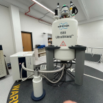
Nuclear Magnetic Resonance: Bruker AVII 600 MHz NMR Spectrometer

The AVII 600 MHz NMR spectrometer is equipped with cryoprobe and is utilized for experiments where the upmost of sensitivity is required, such as protein studies and natural product characterisations.

Bruker AVIIIHD 600 MHz NMR Spectrometer

Used for

Characterisation of natural products, protein NMR structure determination. Variable temperature work is not permitted.

Specifications

  • 5 mm TXI Cryoprobe with Z-gradient (1H/13C/15N/2H observe)
  • 3 high power channel operation
  • SampleXpress Lite 16 Holder Autosampler
  • BCU05 (Bruker Chiller Unit)

Contact

Remi Rouquette
4 Wally’s walk, room 221
Department of Molecular Sciences
Macquarie University
remi.rouquette@mq.edu.au
T: 02 9850 4219

Rates

Discounted rates apply for Macquarie university researchers.

Location

4 Wally’s walk, room 014 (highly restricted access)

Bookings

Facility homepage

Raman

Used for

High Precision Isotope Ratios

Contact

Peter Wieland
E5B - 10 Sir Christopher Ondaatje Avenue, Room 206 
Peter.Wieland@mq.edu.au
T: 02 9850 4402

Location

E5B - 10 Sir Christopher Ondaatje Avenue, Room 135

Spectrofluorometer: Jasco FP-8500

The FP-8500 is a spectrofluorometer for measurement of steady-state fluorescence and luminescence, emission and excitation, across a wavelength range of 200 to 750 nm. The FP-8500 has high resolution and standard automatic band-pass filters to obtain spectra without the artificial peaks due to second order diffracted light. The FP-8500 is also fitted with shutters to protect samples when not being measured and rejection filters to minimise stray light. A Peltier temperature control unit is also available for temperature controlled measurements, making this instrument ideally suited for kinetics and thermodynamics based experiments.

Specifications

  • Light Source: High intensity continuous output Xenon arc lamp
  • Wavelength range: 200 to 750 nm
  • Sensitivity: S/N 75,000:1 (RMS, water Raman)
  • Scanning Speed: 10-60,000 nm/min

Contact

Alison Rodger or Sophie Goodchild
4 Wally’s walk, laboratory 302
Department of Molecular Sciences
Macquarie University
sophie.goodchild@mq.edu.au
T: 02 9850 8235

Rates

Discounted rates apply for Macquarie university researchers.

Location

4 Wally’s walk, room 302 (restricted access)

Bookings

Facility homepage

UV-visible : Jasco V-760

The V-760 is a double-beam UV-Visible spectrometer that measures light absorption, reflectance or transmittance in the visible to ultraviolet region (187-900 nm).  The extended photometric linearity of the V-760 (up to 6 AU) enables measurement of highly absorbing samples which is particularly useful for materials with low transmission properties such as solid films. The V-760 is also fitted with a 6-position multi-cell changer and Peltier temperature control unit making it an ideal UV-vis instrument for kinetic and thermodynamic experiments.

Specifications

  • Optical System: Czerny-Turner mount, double monochromator, fully symmetrical double beam
  • Light Source: Halogen lamp, deuterium lamp
  • Wavelength range: 187 to 900 nm (+/-0.1 nm at 656.1 nm)
  • Photometric Range: -4-6 AU +/-0.3 %T
  • Scanning Speed: 10-400 nm/min

Contact

Alison Rodger or Sophie Goodchild
4 Wally’s walk, laboratory 302
Department of Molecular Sciences
Macquarie University
sophie.goodchild@mq.edu.au
T: 02 9850 8235

Rates

Discounted rates apply for Macquarie university researchers.

Location

4 Wally’s walk, room 302 (restricted access)

Bookings

Facility homepage

X-ray fluorescence: Bruker M4 Tornado Micro XRF

Used for

In-situ major element composition and elemental distribution of whole samples

Specifications

  • Typical Sample Type: Pretty much anything that will fit inside and withstand vacuum
  • Chamber size: 600mm x 360mm x 260mm
  • Resolution down to 20 μm

Contact

Timothy Murphy
E5B - 10 Sir Christopher Ondaatje Avenue, Room 204
Timothy.D.Murphy@mq.edu.au
T: 02 9850 8258

Location

E5B - 10 Sir Christopher Ondaatje Avenue, Room 206

X-ray fluorescence: Panalytical Axios XRF Spectrometer

Used for

Major element analysis of fused glass beads produced from rock powders

Specifications

  • Typical Sample Type: 1 gram of <63 μm rock powder
  • Fused glass beads produced in-house using your sample
  • 1 kW Source

Contact

Peter Wieland
E5B - 10 Sir Christopher Ondaatje Avenue, Room 206
Peter.Wieland@mq.edu.au
T: 02 9850 4402

Location

E5B - 10 Sir Christopher Ondaatje Avenue, Room 206

Surface Plasmon Resonance (SPR)

Biacore X100 biosensor

Protein-protein and protein-ligand interactions can be studied using surface plasmon resonance. A protein or a ligand is bound to the surface of a sensor chip (purchased by the user from Biacore) using one of several standard chemical methods, e.g. a biotinylated ligand may be bound to a streptavidin chip. Buffer containing the analyte is then allowed to flow over the surface of the chip, with binding and release monitored in real time.

Used for

Studying protein-ligand interaction

Specifications

  • Automatic sampling loading
  • Size limit: > 100 Da
  • Typical concentrations (proteins): pM - μM range
  • Sample volume50-120 μl
  • Flow rate: 1-100 μl/min
  • Temperature: 4 - 45°C

Contact

Remi Rouquette
4 Wally’s walk, room 221
Department of Molecular Sciences
Macquarie University
remi.rouquette@mq.edu.au
T: 02 9850 4219

Location

4 Wally’s walk, room 311 (MUCAB)

Bookings

Facility homepage

Thermal analysis

Isothermal Titration Calorimetry: Microcal Auto iTC200

The MicroCal Auto-iTC200 is a fully automated, low volume, highly sensitive isothermal titration calorimeter. Binding affinity, stoichiometry, enthalpy and entropy are measured in solution in a single experiment, without labeling.

Used for

Used for characterizing molecular interactions of small molecules, proteins, antibodies, nucleic acids, lipids and other biomolecules. Can be used to confirm binding, determine stoichiometry, detect protein-protein or protein-ligand interactions.

Specifications

  • Measures Affinity (Kd), enthalpy (ΔH), entropy (ΔS) and stoichiometry
  • Sample capacity: 4x 96 well plates
  • Sample volume: 370 μL
  • Cell volume: 200 μL
  • Injection syringe volume: 40 μL
  • Temperature range: 2 - 80˚C

Contact

Remi Rouquette
4 Wally’s walk, room 221
Department of Molecular Sciences
Macquarie University
remi.rouquette@mq.edu.au
T: 02 9850 4219

Location

4 Wally’s walk, room 311 (MUCAB)

Bookings - only after induction and training

Facility homepage

TGA- TA instrument Thermal

Thermogravimetric analysis monitors the weight change of a wide range of samples upon variation of temperature

Used for

Mass variation of sample under normal or inert atmosphere.

Specifications

  • Heating up to 1000°C
  • Aluminum or ceramic pans
  • Air or Nitrogen atmosphere

Contact

Remi Rouquette
4 Wally’s walk, room 221
Department of Molecular Sciences
Macquarie University
remi.rouquette@mq.edu.au
T: 02 9850 4219

Rates

Discounted rates apply for Macquarie university researchers.

Location

4 Wally's Walk, room 202.

Bookings

Facility homepage

Back to the top of this page